
WHAT IS A DVT RECORDING?
DVT stands for Dental Volumetric Tomography and is the latest generation of 3D CT devices. It impresses with a very good resolution or recording quality and a low radiation dose.
WHAT IS THE DIFFERENCE BETWEEN CT AND CBCT?
CTs are the conventional devices for 3-dimensional diagnosis. The earlier models still had a comparably high radiation output, which could be gradually reduced, also thanks to digitization. The latest generation of CT devices are the so-called Cone Beam CT-s (CBCT), which now have a very low impact. The resolution has also gradually improved, helping dentists and maxillofacial surgeons to better analyze the situation and plan. CBCTs are the newest, completely digital, devices of 3D opinion making.
HOW HIGH IS THE RADIATION OF A CBCT RECORDING?
The radiation that a patient receives during a CBCT recording is now very low and in terms of value is about the amount of jet, which is exposed to an hour of flight at 10,000 meters altitude, so no one should have any concerns any longer, because the amount of radiation is completely harmless.
WHAT RESOLUTION HAS A CBCT RECORDING?
A DVT image consists of several hundred X-ray images, from which the volume data sets are calculated by means of mathematical formulas. The latest devices have an optimization function of the X-ray images, which are often distorted, so that a very accurate image can be obtained.
WHEN IS A DVT RECORDING REQUIRED?
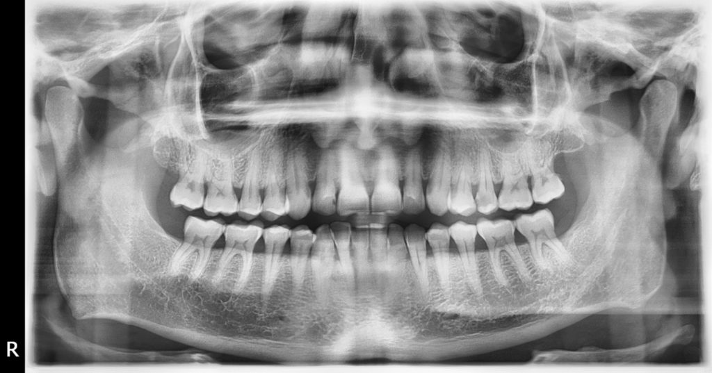
A DVT or CT scan is mostly used in maxillofacial surgery. In some cases, a 3D diagnosis is also required for orthodontic treatments. Prior to orthodontic surgery, an advance 3D diagnosis is mandatory. In any case, prior to an implant surgery, preliminary examinations must take place. First, a panoramic x-ray is taken. When evaluating the X-ray, it is decided which teeth must be replaced. Then it has to be judged whether there is enough bone mass to even place implants. Since the panoramic radiograph is a two-dimensional image showing only height and width, a 3D scan with CT or a volumetric tomograph must be performed to check the bone depth as well. Furthermore, only a CT scan allows an accurate measurement of the bone supply, on which the size of the implants to be used depends. In addition to the bone mass, the general state of health of the patient also plays a decisive role in determining whether appropriate treatment is possible. Circumstances that greatly affect the success of implant placement are e.g. Diabetes or heavy smoking.
Planning: If this phase is completed successfully and implant treatment is possible, then the new teeth need to be planned on the dental implants. The solution decides how many dental implants are needed, which dimensions are necessary and whether an interim care for the healing time should be made. During this planning the exact treatment plan, the timing and the exact costs can be determined.
The dental implants are placed under sterile conditions, mostly in local anaesthesia. For anxiety patients or in case of large surgeries the treatment can also take place under general anaesthesia.
Procedure: After a small incision in the mucosa, the bone surface is exposed, formed the cooking nest of the dental implants and then they are used.
Immediately after the treatment, a panoramic x-ray is taken, which is absolutely necessary for conscientious implant treatment. After about a week, the sutures are removed or the oral surgeon uses self-dissolving threads. The healing phase usually lasts three months, although if bone augmentation or sinus lift were made, the healing time can also take six, in extreme cases, up to nine months. This time is necessary for the bone cells to grow directly into the micropores on the implant surface. This bone cement growth (osseointegration) ensures the stable attachment of the dental implants, after which they can withstand the later, permanent masticatory pressure without damage. Thereafter the preparation of the final teeth takes place. First, the healing implants are uncovered under the palate. This means no more surgery, but only the liberation of the so called closing screw (healing screw) through a small incision in the mucosa and the use of so called palate shaper (gingiva former), which forms the palate in a few days. Thereafter, the impression is taken, on the basis of which, the previously planned teeth are created in the dental laboratory within a short time.
With the finalization of the implant-carried teeth the relationship between the patient and the dentist does not end yet, because the most important prerequisite for the long-term success of implant-carried tooth-replacement is the cleanliness of the mouth and the regular dental check. A check-up should be performed at least once a year, with the dentist checking the condition of the tooth replacement, as well as the attachment of bones and rod around the implants, and making corrections if necessary. Also a thorough professional cleaning is necessary.
Planning bone augmentation or sinus lift
The surgeon first measures the dimensions of the bone and if this is inadequate, bone augmentation or sinus floor elevation (raising the maxillary sinus) must be done. Depending on the situation, this can be done six months before, or together with the implantation. The implant screws must be completely surrounded by the bone, laterally at least 2-3 millimetres, oblong ideally 10-12 millimetres of bone needs to be present. If this is not the case, then the bone is built up with bone substitute material (mostly made of cattle). Sometimes also the own bone of the patient or the combination of the own bone and bone substitute material is being used.
Planning implants
The implants are planned after bone measurement or after a successful bone graft on the 3D image. The oral surgeon defines the positions of the implants and consequently the size of the implants to be used.
Planning implant template
Often a template for implant insertion is also used. This is digitally planned in advance based on an impression and the CT or DVT images and is produced by means of 3D printing. The advantage of the template is that the position of the implants is already physically pre-defined, the maxillofacial surgeon “only” has to perform the implants through the holes of the template so that the implants get in the correct position. The template has the following advantages:
– faster treatment
– Safe treatment
– Minimally invasive treatment
For surgical extraction or wisdom tooth removal
Even with complicated tooth removal, especially when removing wisdom teeth, a tooth CT scan may be necessary, because not infrequently these teeth have 4 or 5 roots whose position and shape cannot always be determined clearly based on the 2D image.
WHAT IS THE COST OF A CBCT TAKE?
A CBCT or DVT take usually costs between £200 and £400 € in the UK. At Evergreen Dental the price is 110 € (£95).
CAN AN EXISTING CT SCAN BE EVALUATED BY EVERGREEN DENTAL?
Yes, this is absolutely possible if the DVT or CT recording is saved on an external medium. CT or DVT recordings contain a large amount of data of up to several gigabytes and must therefore be stored either on a CD-ROM or DVD disc. Since the images can be controlled manually and there are several important functions, such as the measurement, the CT and DVT that need to be used, diagnoses always work only with the associated software that must be installed on the user’s PC therefore, most of the time the installation software is also stored on the same disk as the recordings themselves. Using the DVD player of the computer the software can be installed and then the appropriate recording will be opened. The software and the recorded images can also be stored on a cloud server or sent via a large file transmitter, so that fast access, even from a distance, should be possible.
DO YOU HAVE MISSING TEETH?
In this case, we advise you to come to Budapest for a preliminary examination for only 199 € including travel expenses and to have a 3D image taken at the same time in order to get a perfect picture of the situation and the necessary treatment. The preliminary examination can usually also be combined with the treatment.